Genome Medicine | Single-cell genomic and transcriptomic landscapes of primary and metastatic colorectal cancer tumors
Colorectal cancer (CRC) is one of the most common cancers worldwide, which has high mortality rate. Metastasis of colorectal cancer is one of main causes of its death. In recent years, high-throughput sequencing technology has been widely used to study the pathogenesis and molecular mechanism of several cancers, including CRC. However, the intratumoral heterogeneity and relationships among different omics of CRC have not been systematically investigated. Studies that analyzed the subclonal composition and metastasis models of CRC are still unmet. To fill the gap, Tang Lab from Biomedical Pioneering Innovation Center (BIOPIC) collaborating with Wei Fu’s team from Peking University Third Hospital, recently published a paper entitled Single-cell Genomic and Transcriptomic Landscapes of Primary and Metastatic Colorectal Cancer Tumors on Genome Medicine on July 16th, 2022. This study not only performed a single-cell transcriptomic survey, but also whole genome sequencing (WGS) and multi-region whole exome sequencing (WES) at bulk level of metastatic CRC patients to investigate multi-omics changes during tumorigenesis of CRC.
In this study, researchers collected 11 adjacent nontumor colonic mucosa (N), 9 primary tumors (PTs), 12 matching lymph node metastases (LyMs), 7 liver metastases (LMs), 3 omentum metastases (OMs) and 1 liver normal tissue (LN) form 11 patients mCRC patients. A total of 8,085 cells single cells were retained after the stringent filtration and used for subsequent transcriptome analysis. In addition, the study also performed whole genome sequencing of 84 samples from 9 patients and whole exome sequencing of 81 samples to systematically analyzed the characteristic of mCRC.
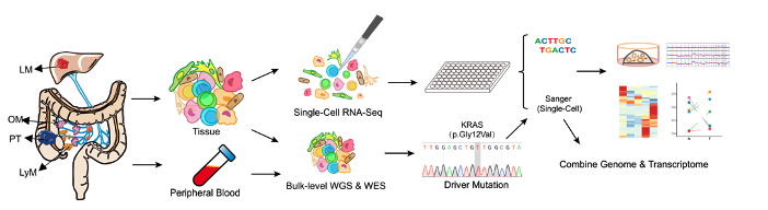
Fig1 The workflow of this research
The study had the following key findings:
SOX9/MKI67 -positive cells may have potential of self-renewal and differentiation.
By systematically compared the cell composition of tumor tissues and adjacent normal tissues, the study found that the proportion of T cells and fibroblasts in tumor tissue increased significantly, and the marker genes of Paneth cell (LYZ+ cell) and intestinal epithelial stem cells (SOX9+ and MKI67+ cell) were significantly increased. The adjacent normal tissues were mainly composed of differentiated mature epithelial cells, such as enterocyte and goblet cells. By establishing tumor organoids and comparing the proportions of differentiated cells and stem cells in tumor organoids after short- and long-term culture, the study found that the proportion of SOX9+MKI67+ cells was increased significantly compared to tumor cells in vitro (tumor cells in vivo: 24%-26%, versus tumor organoids in vitro: 74%). And the proportion of cells with high expression of differentiated cell characteristic gene CA2 increased significantly after long-term culture of tumor organoids, indicating that SOX9/MKI67 double positive cells might have the potential for self-renewal and differentiation, which are the tumor stem cell-like cell features.
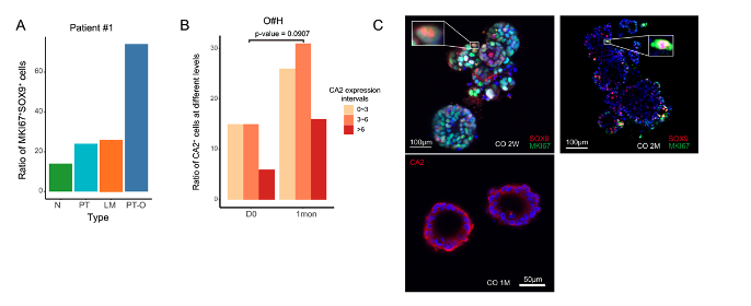
Fig 2 A. Bar plot shows the ratio of SOX9/MKI67 double positive cells of different regions. N, normal tissue; PT, primary tumor; LM, liver metastasis. B. The bar plot shows the ratio of cells that expressed different level of enterocyte marker. C. Immunofluorescence staining of SOX9, MKI67 and CA2 on cultured tumor organoid.
To further validate the differentiation potential of SOX9+MKI67+ double-positive cells, the study performed high-precision single-cell transcriptome sequencing analysis of five organoid spheres in a patient. The cells in an individual organoid sphere were theoretically just derived from a few original cells, or even from a single cell. Combined with single-cell mitochondrial mutation analysis, the researchers traced the origin of cells to infer the differentiation path. Results showed that cell clones with the same mitochondrial mutation in a single organoid sphere co-exist the stem-like cells and differentially mature cells. which further verified that SOX9+MKI67+ double positive cells were tumor stem cell-like cells with the potential for differentiation and maturity.

Fig 3 A. Organoids culture procedure and the workflow of RNA-seq experiment. B. Mitochondrial mutation of patient #1 derived organoid, including clone composition (top), expression of marker genes of different cell types (bottom)
PPAR signaling pathway were abnormally activated in colorectal cancer tumor cells.
By comparing the gene expression characteristics of normal intestinal epithelial cells and tumor cells, the study found the abnormally activation of the PPAR signaling pathway in tumor cells. In order to further explore the role of PPAR signaling pathway on tumor growth and its potential molecular mechanisms, the study constructed colorectal cancer organoid culture system. When the PPAR signaling pathway of tumor organoids was inhibited by small molecule inhibitors, the proliferation rate of tumor cells was significantly decreased, while the apoptosis was significantly increased. More importantly, the tumor killing ability of PPAR signaling pathway inhibitors is comparable to that of the commonly used anti-cancer drug 5FU in the clinically treatment of CRC. It is worth noting that when the PPAR signaling pathway and the WNT signaling pathway were simultaneously inhibited, tumor cells were further killed. Therefore, the researchers further explored the possibility of combining PPAR signaling pathway and WNT signaling pathway inhibitors, and the experimental results showed that the combination of PPAR signaling pathway and WNT signaling pathway inhibitors were dose dependence. Treating cells with low-dose PPAR signaling pathway inhibitors and WNT signaling pathway inhibitors can improve the killing effect on tumors, which has important guiding significance for the clinical treatment of colorectal cancer.

Fig 4 A. Bar plot shows the ratio of apoptotic cells. B. Dot plot showing the proliferation ratio of cells. C. The line plot showing the cell viability of tumor organoid (O#L) under different concentrations of drugs.
Tumor cells with different metastatic sites in the same colorectal cancer patient often have independent origin characteristics.
The study used high-precision single-cell transcriptome data to analyze chromosomal copy number variation (CNV) and mitochondrial point mutations in tumor cell at the single-cell level, and constructed tumor subclonal composition and tumor metastasis patterns in 9 colorectal cancer patients through integrated analysis. The study found that colorectal tumor cells have high-frequency mitochondrial mutations, and that mitochondrial mutations were strongly correlated with CNVs and gene expression patterns. Therefore, these tumor-specific mitochondrial mutations can be used to accurately identify and distinguish normal epithelial cells, tumor cells with insignificant CNVs and tumor cells with obvious CNVs. In addition, the study used CNVs and mitochondrial mutations to verify the possibility of independent origin of lymph node metastatic tumor cells and liver metastatic tumor cells in the same colorectal cancer patient at the single-cell level. (Different tumor subclones in primary tumors were independently transferred to the lymph nodes and liver respectively, rather than the tumor subclones first metastasis to lymph nodes and then metastasis from lymph nodes to the liver). It was also revealed that tumor cells with different distal metastatic sites (e.g., liver metastases and omental metastases) also have the possibility of independent origin.
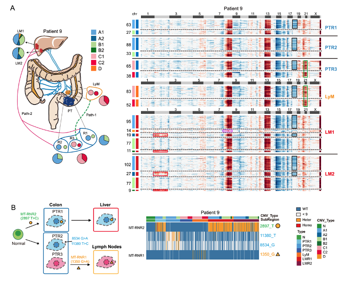
Fig 5 A. CNV pattern and sampling strategy of patient #9. B. The diagram showing the tumor metastasis path of patient #9.
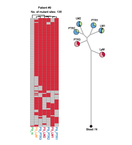
Fig 6 Heatmap showing the reginal distribution of somatic mutations in all samples from patient #9.
EKC/KEOPS complex can promote the metastasis of colorectal cancer tumor cells with TP53 mutation.
By simultaneously performing single-cell transcriptome sequencing of individual cells and cDNA Sanger sequencing of specific driver gene mutations, the study analyzed the relationship between driver gene mutations and gene expression characteristics in tumor tissues at the single-cell level. TP53 mutation is one of the most common genetic mutations in colorectal cancer patients, the study conducted tumor subclonal analysis and tumor metastasis pattern reconstruction in two patients with TP53 gene mutations, and found that the high expression of TP53RK and TPRKB genes in the EKC/KEOPS complex can promote the metastasis of tumor cells with TP53 mutations. Previous studies have shown that the loss of TRPKB expression will cause the growth of TP53 mutant cells to be inhibited, while TP53 wild-type tumor cells are almost unaffected by the loss of TPRKB expression, indicating that TRPKB has an important regulatory role on TP53 mutant cells. The study validated the important role of the EKC/KEOPS complex on tumor metastasis with in vivo tumor cells within single individuals, which could large eliminate the influence of irrelevant interfering factors, such as growth environment and individual differences.

Fig 7 Workflow of single-cell transcriptome sequencing and cDNA Sanger sequencing of specific driver gene mutations.
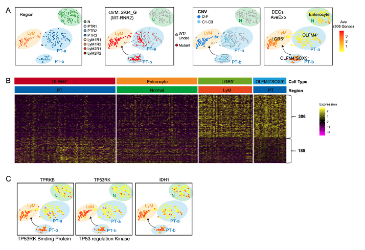
Fig 8 A. The tSNE map of patient #4, which has a TP53 frame-shift mutation. B. Heatmap shows the expression pattern of DEGs between OLFM4+ SOX9+, and OLFM4+ in all four cell groups of patient #4. C. The expression levels of IDH1, TPRKB (TP53RK-binding protein), and TP53RK (TP53 regulation kinase) were projected on tSNE maps.
In summary, through single-cell multi-omics analysis, the study explored the dynamic changes of cell type composition and the key signaling pathways expression during the progression and metastasis of advanced colorectal cancer, and the relevant conclusions were systematically verified by the in vitro organoid culture system. At the same time, the organoid culture system provides a potential drug combination testing strategy for the treatment of colorectal cancer. In addition, the study deeply studied the subclonal composition of primary tumors and tumors with different metastatic positions, unrevealed the tumor metastasis features, explored the relationship between driving gene mutations and gene expression, validated the important role of driving gene mutations on tumor metastasis at the single-cell level.
Dr. Rui Wang, Dr. Jingyun Li, Dr. Yunuo Mao from Peking University, Dr. Xin Zhou, Dr. Wendong Wang from Peking University Third Hospital, Dr. Shuai Gao from China Agricultural University are the co-first authors of the paper. Professor Fuchou Tang from Beijing Advanced Innovation Center for Genomics, Peking University and Professor Fu Wei from Department of General Surgery, Peking University Third Hospital are corresponding authors.
Link:
https://genomemedicine.biomedcentral.com/articles/10.1186/s13073-022-01093-z