GPB | Single-cell sequencing reveals clearance of blastula chromosomal mosaicism in in vitro fertilization babies
On August 6th, 2022, Professor Fuchou Tang’s group in Biomedical Pioneering Innovation Center (BIOPIC) and Beijing Advanced Innovation Center for Genomics (ICG) in Peking University, collaborating with Professor Yuanqing Yao’s team in Chinese PLA General Hospital, published an article entitled “Single-cell sequencing reveals clearance of blastula chromosomal mosaicism in In Vitro fertilization babies” on Genomics, Proteomics & Bioinformatics (GPB).
Embryo mosaicism is defined as the coexistence of normal diploid cells and abnormal aneuploid cells in the same embryo, which is usually caused by chromosome segregation errors in some embryonic cells during mitosis. Previous studies have shown a high rate of mosaicism in human preimplantation embryos, ranging from 3% to 26% in trophectoderm (TE) biopsies [1]
Despite that mosaic embryos may fail to implant or suffer miscarriage after implantation, many of them are able to develop to live birth. Pregnancy and live birth rates after mosaic embryo transfer vary from 16% to 47% [1-3]. However, postnatal evaluation of transferred mosaic embryos is currently insufficient. Particularly, data on postnatal chromosomal testing at single-cell resolution are still lacking.
In this study, the authors analyzed seven healthy infants born from mosaic blastocyst transfer. The peripheral blood mononuclear cells were collected for single-cell multi-omics sequencing, and the identity and chromosome copy number of individual cells were analyzed. The authors explored the chromosomal copy number status of peripheral blood mononuclear cells at single-cell resolution, and assessed whether these healthy infants also had low frequency of the same chromosomal copy number alterations (CNAs) identified in the TE biopsy (Figure 1).
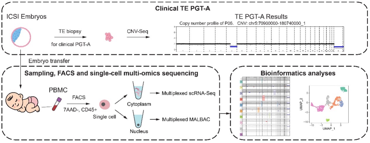
Figure 1 Study design
The study found that:
Early embryonic chromosome copy number alterations were not detected in peripheral blood monocytes of all seven infants. Single-cell multi-omics sequencing was performed on a total of 1616 cells from seven in vitro fertilization (IVF) babies derived from mosaic embryos and three IVF babies from normal euploid embryos (as controls), with an average of 100 to 300 single cells per baby. This study focused on chromosomal regions in which mosaic embryos had copy number alterations at trophectoderm biopsy. The results showed that the peripheral blood mononuclear cells of these IVF babies had normal copy numbers in these corresponding chromosomal regions (Figure 2). One exception was a cell from case P05, which showed partial deletion in the abnormal region in the trophectoderm biopsy; however, the length of deletion was much shorter than that in the original region, and the ploidy change was opposite-gained copies in TE biopsies, whereas lost copies in peripheral blood mononuclear cells were detected. Therefore, the study found that the chromosomal copy number abnormalities occurring in the early embryo were no longer detectable in peripheral blood monocytes from IVF babies derived from mosaic embryos (Figure 2).

Figure 2: Comparison of chromosome copy number alterations between blastocyst trophoectoderm biopsy results and peripheral blood monocytes from IVF babies
A small fraction of peripheral blood monocytes from IVF babies with mosaic embryos had copy number abnormalities in other chromosomal regions. Chromosome copy number alterations (>10Mb in length) still occurred in a subset of peripheral blood monocytes from IVF babies derived from mosaic embryos and control babies derived from euploid embryos, with an average frequency of 1.2% (from 0% to 2.5%) (Figure 3). Aberrant chromosomal regions in these peripheral blood monocytes showed a relatively random pattern. At the same time, the corresponding single-cell transcriptome information also showed that these cells with abnormal chromosome copy numbers were present in various cell types of peripheral blood monocytes (Figure 3). Recently, Yanyi Huang’s group and Jianbin Wang’s group reported that chromosome copy number alterations are common in peripheral blood monocytes of healthy people, and the proportion of abnormal chromosome copy number cells in peripheral blood monocytes is about 7.5% [5]. These 1- to 3-year-old infants appear to have a slightly lower proportion of cells with abnormal chromosome copy numbers than adults.
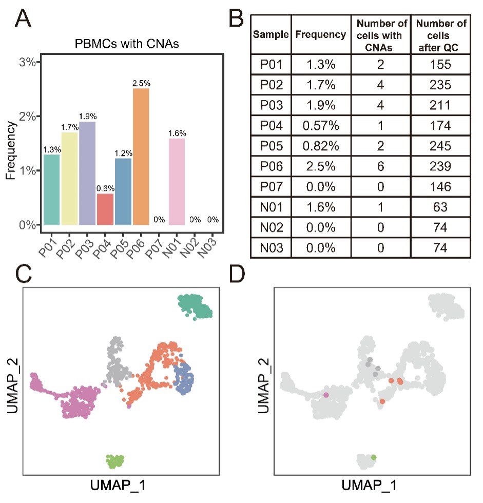
Figure 3: Proportion and distribution of abnormal chromosome copy number cells in peripheral blood monocytes from IVF babies
The above results suggest that the embryonic chromosomal mosaicism problem of these IVF babies is corrected by some mechanisms. There may be two possible scenarios: abnormal aneuploid embryo cells proliferate more slowly than normal euploid cells, or abnormal aneuploid embryonic cells are more prone to apoptosis, and eventually abnormal aneuploid cells are selectively eliminated in mosaic embryos. Studies have shown that aneuploid cells of the inner cell mass lineage of the mouse blastocyst are selectively eliminated by apoptosis, and the proportion of aneuploid cells gradually decreases from the blastocyst stage [6]. Another study showed that the kinetics of proliferation and death of euploid and aneuploid cells are different in early embryos, with aneuploid cells proliferating slowly and prone to apoptosis, whereas euploid cells proliferating quickly and apoptosis rarely occurs [7]. These questions are well worth further exploration in the future.
Taken together, this study provides the first in-depth assessment of IVF babies derived from mosaic embryos from a multi-omics perspective at single-cell resolution, showing that these mosaic embryos can successfully develop into healthy babies with complete correction of embryonic chromosomal mosaicism. This provides new insights into mosaic embryos diagnosed by trophectoderm biopsy. In cases where normal euploid embryos are not available, transfer of mosaic embryos may be considered. This offers new hope for patients with poor ovarian reserve, a limited number of embryos or only embryos with chromosomal abnormalities detected, which represent the majority of IVF patients.
Ph.D. candidate Yuan Gao, Ph.D. candidate Jinning Zhang, Ph.D. candidate Zhenyu Liu and Ph.D. candidate Shuyue Qi are the co-first authors of the paper. Prof. Fuchou Tang and Prof. Yuanqing Yao are the co-corresponding authors of the paper. The project was funded by the National Natural Science Foundation of China, National Key R&D Program of China, the Beijing Advanced Innovation Centre for Genomics at Peking University.
Article link: https://www.sciencedirect.com/science/article/pii/S1672022922000882
References:
[1] Practice C, Genetic Counseling Professional Group of the American Society for Reproductive Medicine. Electronic address aao. Clinical management of mosaic results from preimplantation genetic testing for aneuploidy (PGT-A) of blastocysts: a committee opinion. Fertil Steril 2020;114:246-54.
[2] Greco E, Minasi MG, Fiorentino F. Healthy Babies after Intrauterine Transfer of Mosaic Aneuploid Blastocysts. N Engl J Med 2015;373:2089-90.
[3] Victor AR, Tyndall JC, Brake AJ, Lepkowsky LT, Murphy AE, Griffin DK, et al. One hundred mosaic embryos transferred prospectively in a single clinic: exploring when and why they result in healthy pregnancies. Fertil Steril 2019;111:280-93.
[4] Capalbo A, Poli M, Rienzi L, Girardi L, Patassini C, Fabiani M, et al. Mosaic human preimplantation embryos and their developmental potential in a prospective, non-selection clinical trial. Am J Hum Genet 2021;108:2238-47.
[5] Liu L, Chen H, Sun C, Zhang JY, Wang JC, Du MJ, et al. Low-frequency somatic copy number alterations in normal human lymphocytes revealed by large-scale single-cell whole-genome profiling. Genome Research 2022;32:44-54.
[6] Bolton H, Graham SJL, Van der Aa N, Kumar P, Theunis K, Fernandez Gallardo E, et al. Mouse model of chromosome mosaicism reveals lineage-specific depletion of aneuploid cells and normal developmental potential. Nat Commun 2016;7:11165.
[7] Liu YL, Yu TN, Wang PH, Tzeng CR, Chen CH, Chen CH. Could PGT-A pick up true abnormalities that have clinical relevance? Retrospective analysis of 1043 embryos. Taiwan J Obstet Gynecol 2020;59:496-501.